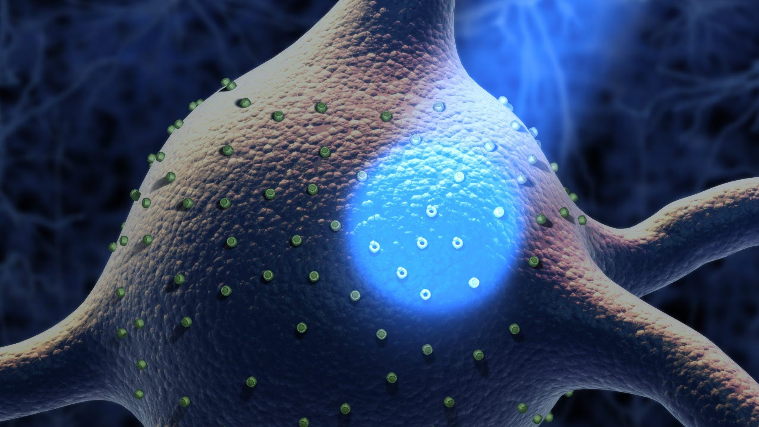
On October 5, 2026, Karolinska Institutet in Stockholm awarded the Nobel Prize for Physiology or Medicine to Karl Deisseroth, Peter Hegemann and Georg Nagelfor the research that allowed the development of theoptogeneticsa technique that has revolutionized the way we study the brain and nervous circuits.
Thanks to optogenetics, exploiting light and particular photosensitive proteins called opsinsresearchers are now able to turn on and off specific populations of neurons in real timeboth in vitro and inside the brains of living animals, and to precisely study the emotions, behavior and the mechanisms underlying many nervous system pathologiesthus shedding light on the properties of the nervous system.
Control cell activity using light
Imagine looking at the sky and wanting to identify a specific star among the hundreds of billions that populate our galaxy, to modify its gravitational field and observe the consequences on all the other stars. It seems like an impossible feat, yet, in a way, optogenetics allows scientists to do something similar: identify with high precision specific populations of cells and change their activity in real time. All this by mainly exploiting two elements: proteins sensitive to light, called opsins (from the ancient Greek ὄψις, meaning sight), and a light source.
But what does it mean exactly optogenetics? To understand this, we can use the words of Karl DeisserothStanford professor and one of the pioneers of optogenetics, who was awarded the Nobel Prize in Physiology or Medicine on October 5, 2026, together with Peter Hegemann and Georg Nagel, precisely for the discoveries that led to the development of this technology. Deisseroth defined optogenetics as:
«the integration of optical and genetic techniques in order to obtain a loss or gain of function of well-defined events in specific cells or specific tissues in vivo».
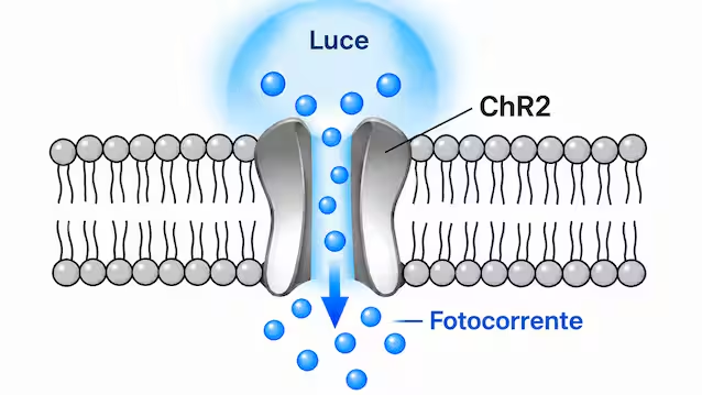
Optogenetics, in fact, exploits one unique property of opsins: when hit by a light source with a specific wavelength, these proteins change their shape, allowing the passage of ions (i.e., of electrically charged particles) through the membrane of the cells that express them, consequently generating an electric current (precisely called photocurrent). The system works similarly to those lamps equipped with a solar panel, which turn on when the sensor detects changes in light. In optogenetics, opsins serve as light sensorsallowing you to control the activity of cells object of study.
A switch to turn neurons on and off
In reality, the photosensitive properties opsins have been known for more than 50 years, since, in 1971, it was discovered that a small protein extracted from microorganisms, called bacteriorhodopsinwas able to transport ions across the cell membrane in response to light. Yet, it was only in 2005 that Professor Deisseroth’s research group managed to demonstrate how to exploit this characteristic to manipulate the activity of live neurons.
Through a study published in the prestigious journal Nature NeuroscienceDeisseroth’s group demonstrated that a particular opsin produced by algae, called channelrhodopsinonce introduced into some neurons in vitro through genetic engineering techniques, was able to make them sensitive to light, allowing the passage of a current through the cell membrane. And, since electrical activity is the basis of the functioning of neurons, this meant having found a sort of artificial switch with which to control the activity of nerve cells. Subsequently, it was demonstrated that this methodology worked not only on neurons in a test tube, but also in an intact brain and in live, free-moving animals, in which the opsins had been expressed in the neurons.
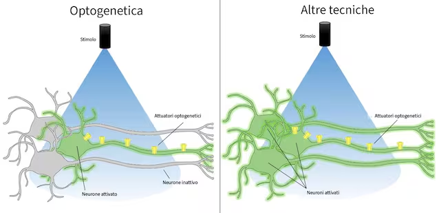
This is the true power of optogenetics in neuroscience: the ability to turn the activity of neurons on or off with one spatial and temporal precision (in the order of milliseconds) difficult to achieve with other techniques, without damage cells or excessively alter their physiology.
Because optogenetics has revolutionized the way we study the brain
Optogenetics was one of those inventions that marked a before and after in our ability to study and understand the brain and neuronal circuits. But how is this possible?
Any function of our brain depends on the coordination of billions of neurons, organized in populations and circuits distributed in the different brain areas which, just like the pieces of a mosaic contribute to forming an image, collaborate with each other, each carrying out a specific role, allowing us to carry out a specific function.
Thanks to genetic engineering techniques, it is possible to choose (even in vivo) the neurons in which to express the opsins, which thus become sensitive to light. In this way, optogenetics allows you to study the properties of nerve cells and manipulate neural circuits with sartorial precision, isolating the contribution of particular neurons compared to all the others. All through a simple beam of light with precise characteristics (manipulated by scientists) and in real time, while, for example, a laboratory animal moves, carries out a memory test or carries out specific behaviors, allowing the consequences of the manipulation on behavior to be immediately observed.
It is precisely this possibility of establishing a cause-effect relationship between the activity of specific neuronal populations and a behavioran emotion or brain function that has transformed optogenetics into one of the most important tools of modern neuroscience.
In just 20 years, this technique has made it possible to better understand the mechanisms underlying some pathologies of the nervous system, such asstroke or Parkinson’s diseaseand to study in depth the role of specific neural circuits involved in memory, emotions and movement control.
Some recent research hypothesizes a possible therapeutic application in humans too, not so much for brain pathologies, where the invasiveness of the technique currently limits its clinical application, but for one of the regions of our body made up of more easily accessible neurons: the retina. Promising studies like “Optogenetic Therapy With UGX-201 in Advanced Nonsyndromic Retinitis Pigmentosa: Safety and Efficacy From an Exploratory Clinical Trial”, they have in fact demonstrated how this technique could be used for partially restore vision in people suffering from retinal degenerationthus identifying one of the first possible therapeutic applications of optogenetics in humans.
Sources:
Deisseroth K., Optogenetics, 2011 The Noble Prize in Physiology or Medicine Boyden ES et al., Millisecond-timescale, genetically targeted optical control of neural activity, 2005 Chen W. et al., The Roles of Optogenetics and Technology in Neurobiology: A Review, 2022 Sahel J.-A. et al., Partial recovery of visual function in a blind patient after optogenetic therapy, 2021 Han