This simple experiment reveals one of the most fascinating mechanisms of the human visual system: thenegative afterimageor afterimage. As you keep your eyes still on the cross, the two halves of your retina are flooded with different colors, red on the left and green on the right. The photoreceptors responsible for those colors progressively tire, reducing their sensitivity. When you move your gaze to the yellow circles, those same “exhausted” areas of the retina are no longer able to respond normally, and the brain makes you see the yellow circles with altered colors.
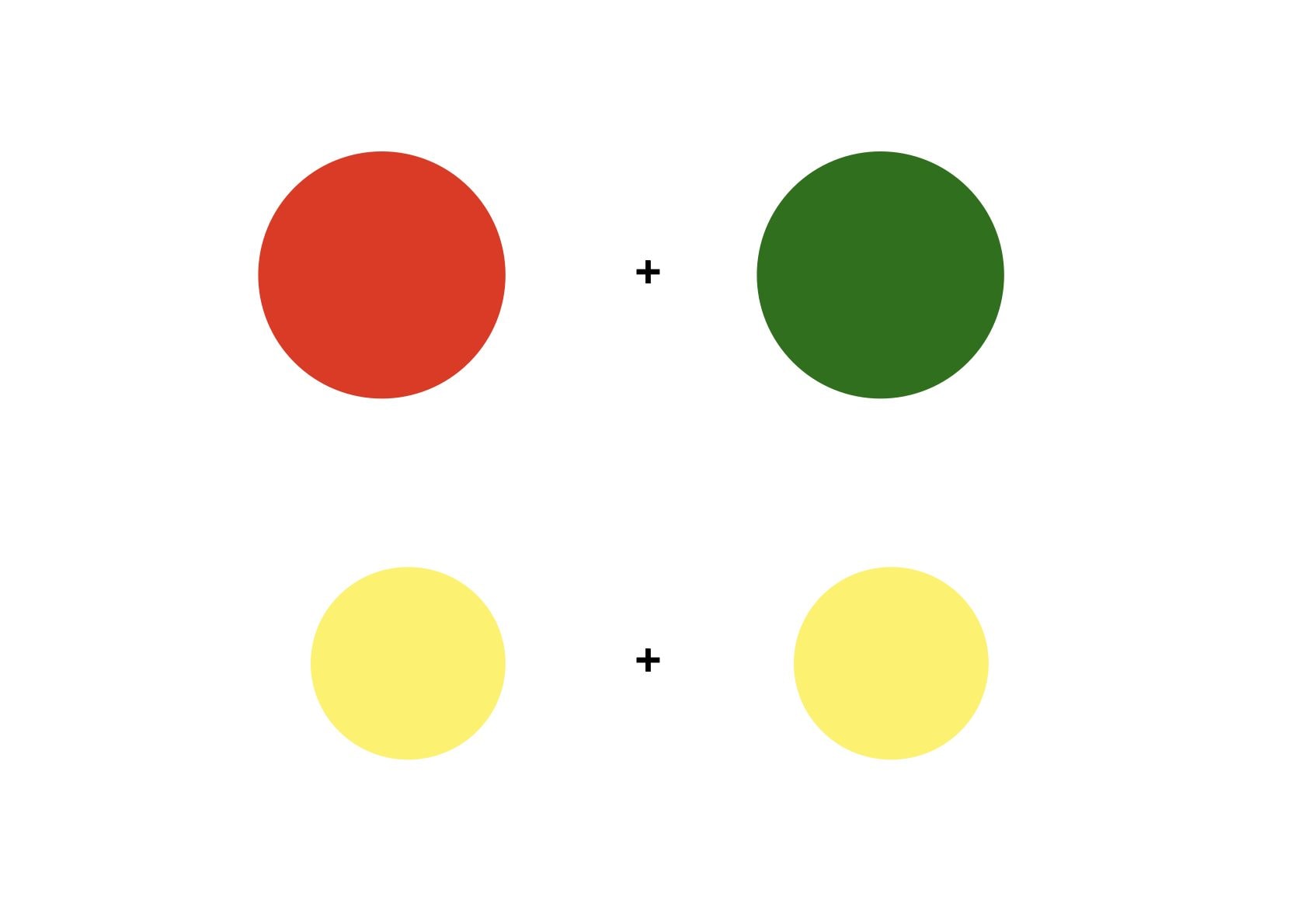
The eye experiment
Attach the cross at the top in the center between the red circle and the green circle to 30 seconds. Then lower your gaze and stare at the cross at the bottom between the two yellow circles. If you performed the experiment well, you will see something surprising: the yellow circle a left will seem to tend towards green or cyanwhile that a right will appear tending towards red or orange. Yet they are physically exactly the same color. How is this possible?
The explanation: retina, cones and color vision
Inside the eye, the retina represents the layer of cells that lines the back of the eye and functions as the “film” of our visual system. Two types of photoreceptors are found in the retina: i rodsspecializing in low light vision, ei conesresponsible for color vision.
There are three types of cones, each sensitive to a different wavelength of light:
- Cones THE (Long), sensitive to red
- Cones M (Medium), sensitive to green
- Cones S (Short), sensitive to blue
When light hits these receptors, they send signals to the brain, which combines them and interprets them as colors.
Sensory adaptation: when receptors get “tired”
During the experiment, while you keep your eyes fixed on the cross at the top, the left side of your retina is flooded with light red (from the left circle), and the right side is flooded with light green (from the right circle). This happens because even if you are staring at a central point, the lateral circles continue to stimulate the corresponding areas of the retina.
After 30 seconds of continuous stimulation, the L (red-sensitive) cones on the left side of the retina are sold outshort of photosensitive molecules. Likewise, the M (green-sensitive) cones on the right side are depleted. This process is called sensory adaptation: it’s the same mechanism by which you stop smelling a room after a few minutes, or feeling the pressure of a watch on your wrist.
The theory of opponent processes: the brain reasons through contrasts
When you move your gaze to the cross at the bottom, the yellow circles stimulate the same areas of the retina that were just “fatigued”. And this is where a second, even more elegant mechanism comes into play.
Our brain does not process colors absolutely, but for comparison and opposition. The cone signals are organized into three opposing channels: Red vs Green, Blue vs Yellow, Light vs Dark. Each channel works like a scale: when one side is active, the other is suppressed.
Here’s what happens in the eye during the experiment:
- Left side (where the red was): The L cones are fatigued and firing a weak signal. When yellow light arrives, which normally stimulates both the L and M cones, the M cones respond regularly, but the L cones almost do not respond. The brain receives an unbalanced signal towards green, and interprets the yellow circle as tending towards green or cyan.
- Right side (where there was green): Mirror situation. M cones are sold out. Yellow light is perceived with a predominance of red, and the circle appears reddish or orange.
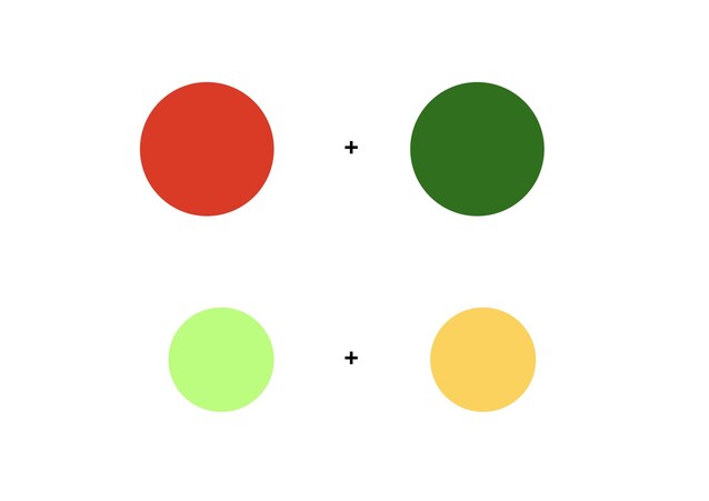
The result is anegative afterimage: each area of the retina projects onto the yellow circle the complementary color of the one it had just fixed. Red leaves a cyan-green ghost, green leaves a red-orange ghost.