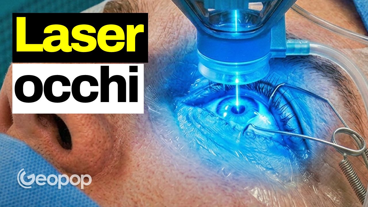
THElaser eye surgery it is an increasingly popular choice for correcting vision defects, allowing you to abandon the use of glasses and contact lenses.
However, many people still have doubts and fears about this type of operation and feel anxious at the idea of operating on such a delicate part of the body. But how much truth is there in these fears?
To respond to these fears, we followed our colleague Alessandra, who underwent refractive surgery at the Vista Vision clinic. We documented the before, during and after the operation to tell you about his experience.
The preliminary phase
Before undergoing the operation, it is necessary to understand whether the operation can be done and, if so, what type of operation. For this reason, specialized doctors must evaluate several parameters. With specific instruments, for example, we inspect the endothelium (the innermost layer of the cornea) or the pressure in the eye. Then we undergo the corneal topographywhich looks at the thickness and curvature of the cornea. Finally, a final test was also performed, theautorefractometrywhich is used to give an estimate of the refraction, therefore of the defect of one’s eyes.
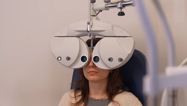
Our colleague’s parameters, as you can see in the video, were also carefully evaluated by the surgeon who will carry out the operation, to understand which type of operation was best suited to her.
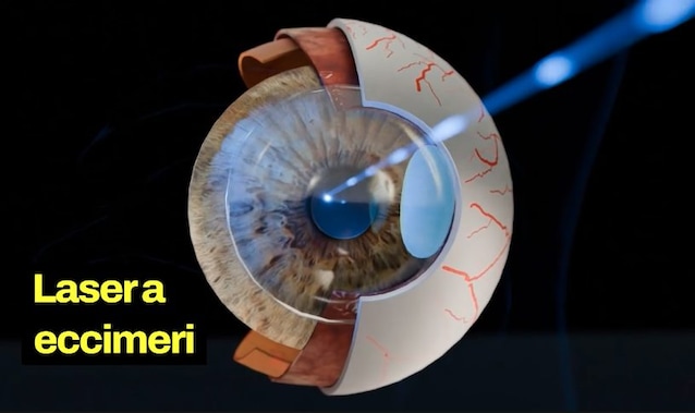
At the end of all these tests, the surgeon opted for the Femtolasik technique, which in practice works like this: a flap of corneal epithelium is created with a femtosecond laser (hence the name), the flap is raised, and then the cornea is shaped with an excimer laser to correct the defect. Finally, the flap repositions itself, and in about 24 hours the vision improves.
The eye operation
On the day of the operation, the surgeon visited our colleague to check the health of her eyes. As you can see from the video, we went down to the operating room floor, where Alessandra lay down on the table.
The last step before the operation is the total anesthetization of the eye with eye drops. After a short period of time, and with the help of special tweezers, the surgeon keeps the first eye open, places the instrument in the appropriate position, and activates the femtosecond laser for only 6 seconds: this step is used to create the corneal flap. The same treatment is also done for the other eye.
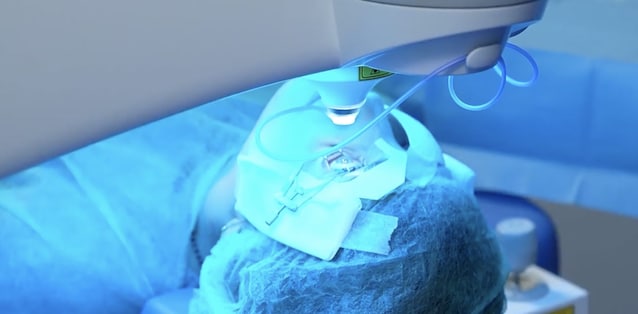
But there is a second step: here the corneal flap is raised, and the excimer laser is activated for 6 seconds. Then the flap is placed again and the same procedure is repeated for the second eye. In the space of just one 15 minutesthe operation is already finished.
The post-intervention
Our colleague initially had blurred vision and a slight headache. Over the next few hours she put on antibiotic drops and experienced some tearing, mild irritation (about 2-3 hours) and mild photophobia, and so the surgeon gave her sunglasses to get through the afternoon.
About 24 hours after the operation Alessandra had one check-up visit to see that everything was healing, and he could see much better than before. Both the next day’s visit and the next week’s visit went well: the eyes were healing perfectly. One thing Alessandra found were the nocturnal haloswhich began to decrease and within 2-3 months, due to a question of neuroadaptation, disappeared.
Ultimately, Alessandra’s experience demonstrates that refractive surgery is not just a medical procedure, but a real investment on your quality of life. Being able to practice outdoor sports without the encumbrance of glasses or waking up in the morning clearly seeing the surrounding world are goals which, thanks to the technologies implemented, as in this case, by companies such as Vision view.