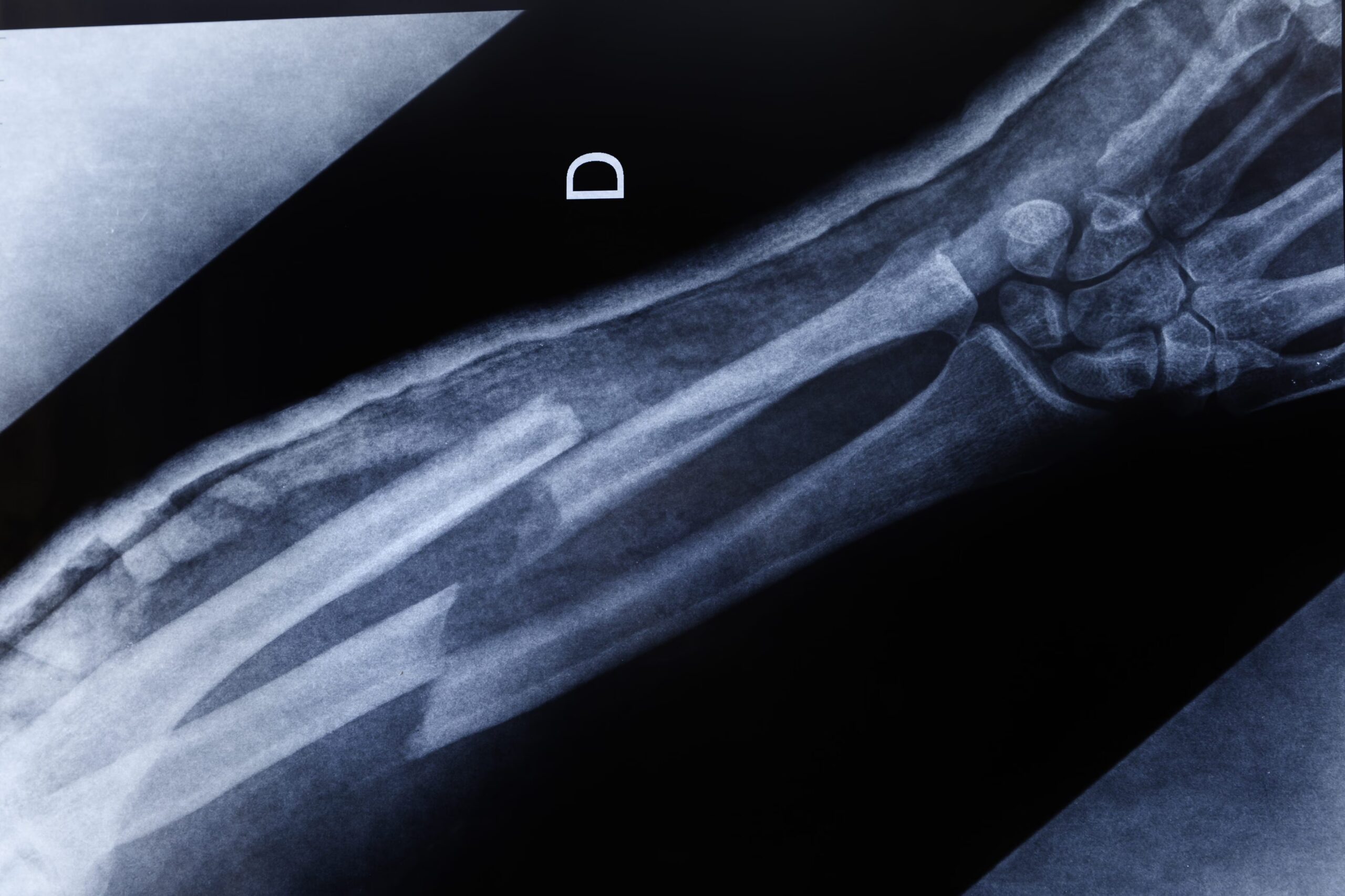
We often imagine bone as a static, immutable tissue, but in reality, like everything in our body, to remain, it must change and constantly changing. Bone tissue is an element dynamic and changingcapable of adapting, repairing itself and with an important role in many physiological functions of the human body, from management of circulating calcium levels to the production, in the marrow, of the precursors of many cell types, as detailed in the dedicated pages on StatPearls of the National Library of Medicine.
Every tissue or organ that, due to a lesion, suffers an interruption of its structure, initiates specific repair processes, to restore its function. Bone is no exception and its repair processes follow an orderly sequence of events starting from lesion of blood vessels and the start of the “construction site” of bone reconstruction. Specialized cells, such as fibroblasts and chondroplasts build an initial “scaffold” made from collagen and cartilagecalled “soft callus“. Only later will the osteoblasts begin to fully reconstruct the bone, forming the “bony callus” definitive. This is why it is fundamental immobilize the limbavoiding excessive loads that would undermine the entire process.
The call to work: the hematoma after the breakup
Bone is a richly vascularized tissue and where a fracture occurs, one also occurs lesion of blood vessels. It will be the blood tissue itself that initiates the repair, through the formation of a hematoma. Blood cells, once they accumulate outside the blood vessels, produce a protein scaffold of fibrin who will act as support and guide for the repair activities. As well documented in the anatomy and physiology manuals of the National Library of Medicine, thanks to the release of various chemical signals, inflammatory cells, endothelial cells and fibroblasts are recruited, together with mesenchymal stem cells, progenitor cells multipotent and not yet differentiated, which will give rise in the subsequent phases to chondroblasts, osteoblasts and osteoclasts, the actors who will actually lay down and remodel the bone tissue.
Inflammationthe presence of many different cells and the initiation of signaling and repair activities make the fracture area a place teeming with activity and for this reason it is also called fracture “hotbed”.. This initial phase is therefore characterized by the mobilization and on-site recruitment of all the necessary actors and takes place already in the first 24 hours.
Early stages of bone regeneration
In the first days after the fracture i macrophages“scavenger” cells capable of engulfing and eliminating both pathogens and cells of the organism, begin to remove all debris from the firebox. At the same time, fibroblasts and chondroblasts, cells that produce cartilaginous connective tissue, a flexible and resistant support tissue, begin to produce collagen fibers and cartilaginous matrix to start tie and reassemble the bone fragments. It is the phase that physiology manuals call “granulation” and which is also explained in detail by the dedicated StatPearls entry, published on the official NCBI (National Center for Biotechnology Information) platform.
The production of real bone, with its resistant mineral part, will take place over time and will exploit the presence of cartilage, more flexible but still resistantto take place on an appropriate basis. This is temporary anchoring structure and support is often called “soft callus“, as opposed to “bony callus“true that will form after the first weeks after the fracture, as it cannot provide structural resistance to loads. Precisely due to the impossibility of supporting loads and the flexibility of the soft callus, in this phase they are crucial bone immobilization systems, such as i chalks.

Towards a more solid structure
After a few weeks the osteoprogenitor cells begin to deposit small three-dimensional units of bone, the bone trabeculae. These resistant, mineralized elements gradually replace the cartilage, stabilizing and rejoining the bone stumps. During the formation of this bony callus, the bone it is not yet able to independently support the full load. In this phase the osteoblasts produce “bone” in abundance, while the osteoclasts reabsorb the excess tissue in the points not subjected to load, shaping the bone exactly based on the mechanical stresses it will have to bear: a principle known as Wolff’s law, also described in the specific section on bone remodeling of the National Library of Medicine, according to which a more stressed bone becomes denser. according to which a more stressed bone becomes denser.
Bone healing is completed with the restoration of the medullary cavity and the complete regeneration of the bone marrow inside. Unlike many other tissues which regenerate partially or to a limited extent after an injury, for example leaving scars, bone is capable of regenerate completelyreturning to having a fabric functionally and structurally identical to that before the injury.