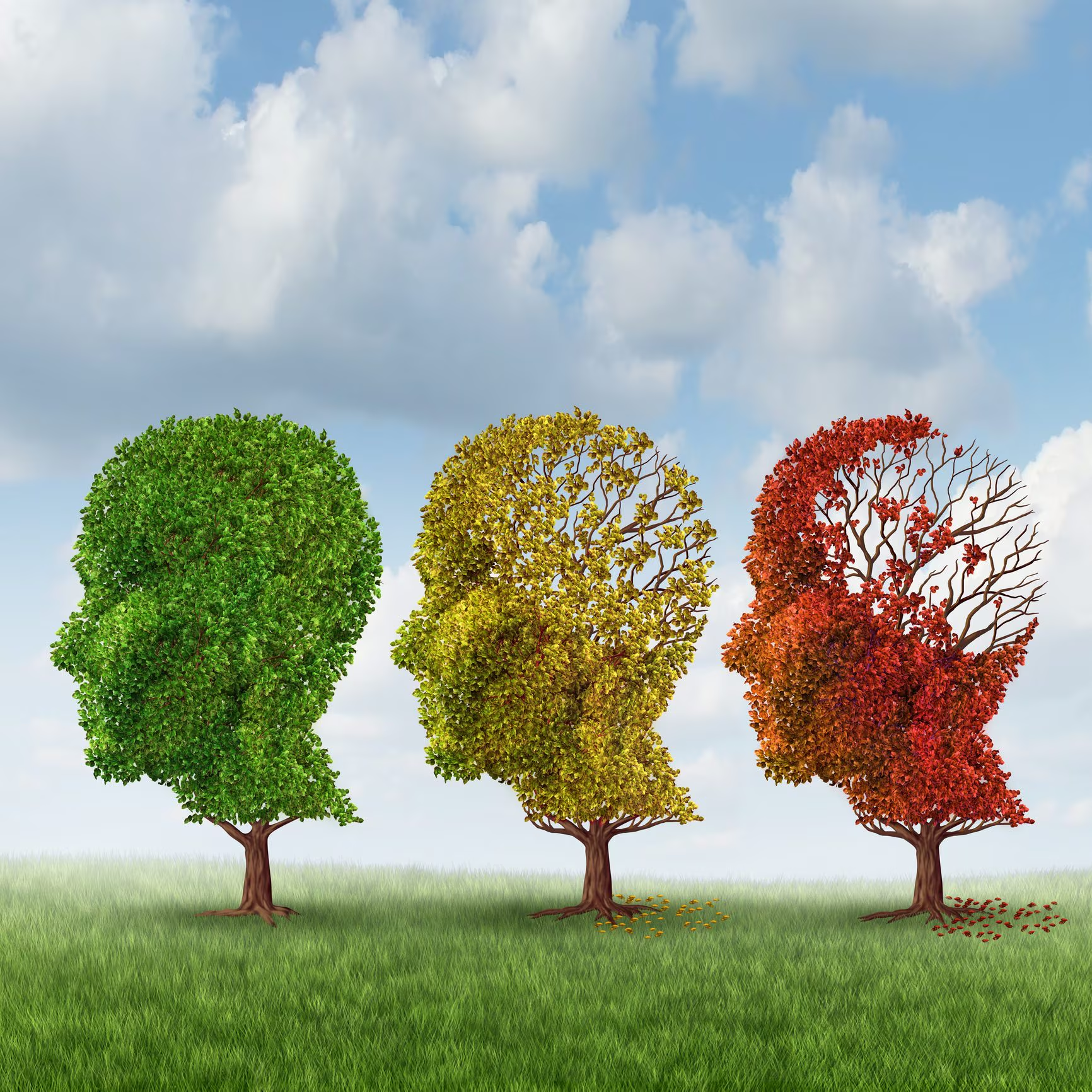
A new study published in Nature from the Icahn School of Medicine at Mount Sinai mapped the development of 1.3 million brain cells from 0 to 97 years old, creating the first molecular map High-resolution image of the long dorsolateral prefrontal cortex the entire span of human life. With this study neuroscience has managed to fill a fundamental gap: Before this work, most studies only analyzed narrow age groups or brains already affected by pathologies, making it impossible to distinguish the physiological changes of normal aging from pathological ones. This study also reveals to us the three phases of brain evolution throughout life and how the functions of the neurons in this area of the brain change with age.
How neurons change during life: the 3 phases and the turning point at 24 years old
We know it, during our whole life the brain evolves considerably. This change reflects some molecular dynamics that occur in the Dorsolateral prefrontal cortexthe site of the brain responsible for reasoning, planning, and emotional control, which is particularly sensitive to development and aging. To understand the scientific causes of this evolution, the study conducted by the PsychAD consortium and the Icahn School of Medicine at Mount Sinai published in Nature analyzed the genetic profile of single brain cells belonging to 284 long healthy donors four broad age groups: development (0 to 19 years), young adults (20-39 years), middle age (40-59 years) and old age (over 60 years). In this way, the researchers discovered that the activity of genes within brain cells does not vary in a linear and constant way over time, but follows a progress in three phases clearly divided.
There first phasewhich spans childhood and adolescence, is characterized by a deep and rapid molecular restructuring. During this period, an intense expression of genes related to synapse formationthe junctions that allow neurons to communicate, the development of neuronal extensions and the construction of brain circuits. This is a time window of extraordinary plasticity, which is key to our ability to learn, but is also particularly sensitive to the genetic predisposition for certain psychiatric disorders. The decisive turning point occurs around 24 years old of age: the analyzes highlight that, at this point in life, the composition of the brain cells stabilizes. So let’s get into it second phasethat of adulthood, marked by a remarkable molecular stability in which gene expression changes very little. Finally, after the age of 60, we enter the third phase of brain aging.
How do brain cells defend themselves and transform as we age?
When it comes to brain agingwe often imagine a progressive and irreparable loss of nerve cells. However, neurons behave differently than we would expect. How do brain cells really react to the passage of time?
The scientific answer that emerged from the study tells us that i principal neurons of the prefrontal cortex demonstrate one extraordinary resilience molecular in healthy aging. These neurons, unlike the cells of the skin, intestine or blood (which are continuously renewed), almost remain the same from our childhood until old age. So what they do is they activate a continuous maintenance which continuously searches for and repairs damage to the double helix of its DNA and accelerates the processes of discarding damaged proteins (the “cellular garbage”).
The true protagonists of change in aging are the glial cellsi.e. the complex network of supporting cells that includes astrocytes, microglia and oligodendrocytes. If in childhood the glial cells work to guide the growth of neurons and isolate the nerve fibers with myelin, in old age they completely reprogram their functions, reorganizing themselves, around the age of 60, to defend the brain tissue. But all this also represents a double-edged sword: the prolonged activation of these immune programs awakens genes associated with the risk of neurodegenerative diseases like Alzheimer’s. Furthermore, the researchers confirmed that these alterations of microglia and oligodendrocytes are mainly concentrated in the white matter adjacent subcortical, the communication highways of the brain, explaining why this very substance is particularly vulnerable to age-related decline.
Why do sleep patterns and the sense of time change in old age?
Many older people experience a lighter and more fragmented sleeptend to wake up at dawn and feel aalteration of one’s internal clock.
To clarify the molecular causes of this phenomenon, the researchers analyzed the diurnal pattern of “clock genes” (the set of genes that regulate circadian rhythms over 24 hours) in the neurons and glial cells of the prefrontal cortex. In young and middle-aged adults, the prefrontal cortex shows perfect temporal orchestration: neurons maintain clearly synchronized circadian rhythms, alternating peaks of activity between day and night to regulate cognitive functions and rest. With the arrival of old age, this neuronal synchrony progressively disappears: Neurons lose the regular oscillation of their clock genes. What is really surprising, however, is that the circadian clock within the prefrontal cortex does not turn off at all in the elderly brain, but changes “manager”.
In fact, microglial cells and oligodendrocytes develop new diurnal rhythms, which are completely absent in youth. These rhythms emerge in glial cells they no longer respond to the alternation of light and dark in the environmentbut they synchronize on the response to stress and the management of damaged proteins. Basically, time management in the elderly brain moves from neurons to microgliatransforming the local biological clock from a tool of cognitive coordination to a temporal mechanism of cellular defense and cleaning.